Microorganisms are too small to be seen with the naked eye, so they must be observed with a microscope. Microorganisms and their structural components are measured in even smaller units, such as micrometer (µm, 10-6), nanometer (nm, 10-9 ) and angstroms (A0 , 10-10).
Microscopy:
Microbiologists employ a variety of light microscopes in their work; bright-field, dark-field, phase contrast and fluorescence microscopes are most commonly used.
The light microscope uses visible light as source of illumination and uses series of glass lenses to form images. Light microscope has magnification of about 1000x and has limit of resolution of about 0.2µm. Both living and dead specimens are viewed with light microscope. The simple light microscope consists of a single lens a magnifying glass. Compound microscopes are made of more than one glass lens in combination. It consists of condenser lens, the objective lens and the eyepiece lens. Condenser lens focus the light from the light source at the specimen. The objective lens is responsible for producing the magnified image. The eyepiece further magnifies the image in combination with objective lens.
Lenses and the bending of light:
To understand how a light microscope operates, one should know something about the way in which lenses bend and focus light to form images. When a ray of light passes from one medium to another, refraction occurs, that is, the ray is bent at the interface. Refractive index is the measure of the relative velocity at which light passes through a material. When light passes from air to glass, a medium with a greater refractive index, it is slowed and bent towards the normal. As light leaves glass and returns to air, a medium with a lower refractive index, it accelerates and is bent away from the normal. As a result the prism bends light passing through it. A lens functions somewhat like a collection of prisms. Light rays from a distant source are focused at the focal point F (Fig. 1). The focal point lies at a distance f, the focal length, from the lens center. Our eyes cannot focus on objects nearer than about 25 cm or 10 inches and can be overcome by using a convex lens as a simple magnifier (or microscope). Lens strength is related to focal length; a lens with a shorter focal length will magnify an object more than a weaker lens having a longer focal length.
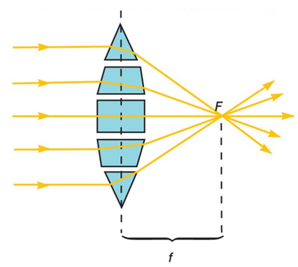
Fig. 1. Light refraction and lens function
Microscopic resolution:
Resolution is the ability of a lens to separate or distinguish between small objects that are close to each other. Much of the optical theory underlying microscope design was developed by the German physicist Ernst Abbe in the 1870's.
d = 0.5λ / nsinθ
d = distance between two objects that reveals them as separate entities
λ = wavelength of light
nsinθ = numerical aperture (NA)
As‘d' becomes smaller, the resolution increases. The wavelength must be shorter than the distance between two objects. Thus greatest resolution is obtained with light of the shortest wavelength, light at the blue end of the visible spectrum (450 to 500nm). The nsinθ or NA is more difficult to understand. The θ = 1/2th angle of the cone of light entering an objective. Light that strikes the specimen after passing through condenser is cone-shaped. Cone of light has a narrow angle then resolution is low. Cone of light has a broad angle and spreads out rapidly after passing through a spectrum, closely packed objects appear widely separated and more resolved. The angle of the cone of light that can enter a lens depends on the refractive index (n) of the medium in which the lens works. The refractive index of air is 1.00, since sinθ cannot be greater than 1.00 (the maximum θ is 90° and sin 90° is 1.00), no lens working in air will have a NA greater than 1.00. The practical way to raise the NA above 1.00 is therefore to increase the refractive index with immersion oil which has a refractive index of glass (1.25-1.4, glass and immersion oil). Air is replaced with immersion oil, light rays that did not enter the objective due to reflection and refraction will now do so and there will be an increase in NA and resolution.
d = (0.5) x (530nm) / 1.25 = 212 nm or 0.2µm
The maximum resolution of a bright field microscope with oil immersion is 0.2µm.