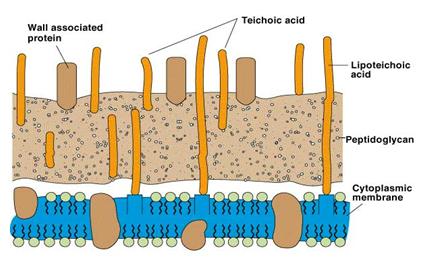
Gram positive cell walls
The cell wall consists of many layers of peptidoglycan, forming a thick, rigid structure, which contains a peptide interbridge. Gram positive cell walls also contain teichoic acids, polymers of glycerol and ribitol joined by phosphate groups. To the glycerol and ribotol groups, the amino acids such as D-alanine or sugars like glucose are attached (Fig. 7). There are two classes to teichoic acids; lipoteichoic acid (if they are attached to the lipid of the plasma membrane) and wall teichoic acid (which extend to the surface of the peptidoglycan and are negatively charged). Teichoic acids are not found in gram-negative bacteria.
 |
|
Some bacteria like Staphylococci and most other gram-positive bacteria have a layer of proteins on the surface of their cell wall peptidoglycan. Some are noncovalently attached by binding to the peptidoglycan, teichoic acids and other receptors and these proteins are involved in the interactions of the cell with its environment. Ex. S-layer proteins. Other surface proteins are covalently attached to the peptidoglycan and in gram positive pathogens these have roles such as aiding in adhesion to host tissues, preventing opsonization and blocking phagocytosis.
Gram negative cell walls
The cell wall contains only a thin layer of peptidoglycan. In E. coli, it is about 2-3nmthick and contains only one or two layers of sheets of peptidoglycan. The peptidoglycan is bounded to lipoproteins in the outer membrane and is embedded in a soft material, the periplasmic gel. Gram negative cell walls do not contain teichoic acids. They are more susceptible to mechanical breakage because of small amount of peptidoglycan. Outer membrane - found primarily in gram-negative cell consists of lipoproteins, liposaccharides and phospholipids and lies outside the thin peptidoglycan layer. The most abundant membrane protein is Braun's lipoprotein, a small lipoprotein covalently joined to the underlying peptidoglycan and embedded in the outer membrane by its hydrophobic end. The outer membrane e and plasma membrane appear to be in direct contact at many locations in the cell wall. The adhesion sites may be regions of direct contact or possibly true membrane fusions. It is proposed that substances can move into the cell through these adhesions sites rather than travelling through the periplasm.