The bacterial cell wall
Is a complex, semi-rigid structure that surrounds the underlying plasma membrane and protects the cell and the internal parts from adverse surrounding environment. Except for Mycoplasmas and some Achaea most bacteria have strong walls that give them shape and protect them from osmotic lysis; wall shape and strength is primarily due to peptidoglycan. The cell walls of pathogenic bacteria have components that contribute to their pathogenicity and protect a cell from toxic substances and are the site of action of several antibiotics. Major functions – prevent cells from rupturing, maintains shape and serves as a point of anchorage for flagella. Christian Gram developed the Gram stain in 1884, and it became evident that bacteria could be divided into two major groups based on their response to staining. Gram positive bacteria stained purple and the gram negative ones colored pink or red by the technique. The gram positive cell wall consists of a single thick layer of 20 to 80 nm of peptidoglycan or murein layer lying outside the cell membrane. In gram negative cells, the peptidoglycan layer is thin of about 2 to 7 nm surrounded by a 7 to 8 nm thick outer membrane. The gram positive cells are stronger than gram negative ones because of the peptidoglycan layer.
A space is seen between the plasma membrane and the outer membrane in electron micrographs of gram negative bacteria and a similar one but smaller gap may be observed between the plasma membrane and wall in gram positive bacteria. This space is called periplasmic space. The substance that occupies the space is the periplasm. The periplasmic space in gram negative bacteria ranges from 1nm to as great as 71 nm. It contains many proteins and participates in nutrient acquisition, hydrolytic enzymes attacking nucleic acids and phosphorylated molecules. The periplasmic space contains enzymes involved in peptidoglycan synthesis and modification of toxic compounds that could harm the cell. Gram positive bacteria may not have a visible periplasmic space but appear to have as many periplasmic proteins and several enzymes that are secreted are often called exozymes.
The Archaea differs from other prokaryotes. Thewalls lack peptidoglycan and are composed of proteins, glycoprotein or polysaccharides.
Structure of Peptidoglycan
The cell wall of bacteria is composed of a macromolecular network of polymer called peptidoglycan (murein). The polymer contains two sugar derivatives, Monosaccharides- N-acetylglucosamine (NAG) and N-acetylmuramicacid (NAM). In addition, several different amino acids, three of which – D-Glutamic acid, D-alanine, and mesodiaminopimelic acid – not found in proteins are present. The pretense of D-amino acids protects against attack by most peptidases. The backbone of this polymer is composed of alternating N-acetylglucosamine and N-acetylmuramic acid residues. A peptide chain of four alternating D- and L- amino acids is connected to the carboxyl group of N-acetylmuramic acid. Many bacteria substitute another diaminoacid, usually L-Lysine, in the third position for meso-diaminopimelic acid. Chains of linked peptidoglycan subunits are joined by cross-links between the peptides. Often the carboxyl group of the terminal D-alanine is connected directly to the amino group of diaminopimelic acid, but a peptide interbridge may be used instead (Fig. 6). Most gram-negative cell wall peptidoglycan lacks the peptide interbridge.
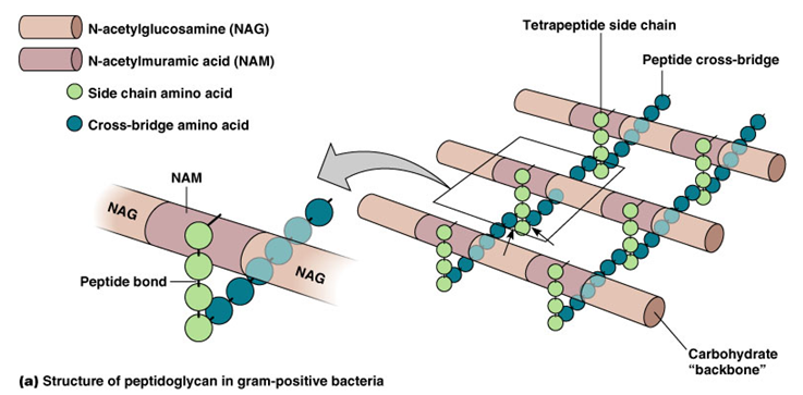 |
Fig. 6. Peptidoglycan structure in gram-positive cell |