| c. Staining: A polychromatic blood stain, such as Giemsa, provides a convenient method of preparing a stained culture. Giemsa stain is usually combined with May–Grunwald stain when staining blood, but not when staining cultured cells. Alone, it stains the nucleus pink or magenta, the nucleoli dark blue, and the cytoplasm pale gray-blue. It stains cells fixed in alcohol or formaldehyde but will not work correctly unless the preparation is completely anhydrous. |
Chromosome Content: Chromosome content or karyotype is one of the most characteristic and best-defined criteria for identifying cell lines and relating them to the species and sex from which they were derived. Chromosome analysis can also distinguish between normal and transformed cells because the chromosome number is more stable in normal cells (except in mice, where the chromosome complement of normal cells can change quite rapidly after explantation into culture).
Chromosome Banding: This group of techniques was devised to enable individual chromosome pairs to be identified when there is little morphological difference between them. For Giemsa banding, the chromosomal proteins are partially digested by crude trypsin, producing a banded appearance on subsequent staining. Trypsinization is not required for quinacrine banding. The banding pattern is characteristic for each chromosome pair.
Other methods for banding are:
- Using trypsin and EDTA rather than trypsin alone
- Q-banding, which stains the cells in 5% (w/v) quinacrine dihydrochloride in 45% acetic acid, followed by rinsing Giemsa banding the slide, and mounting it in deionized water at pH 4.5
- C-banding, which emphasizes the centromeric regions
Techniques have been developed for discriminating between human and mouse chromosomes, principally to aid the karyotypic analysis of human-mouse hybrids. These methods include fluorescent staining with Hoechst 33258, which causes mouse centromeres to fluoresce more brightly than human centromeres.
Chromosome painting: Chromosome paints are available commercially from a number of sources. The hybridization and detection protocols vary with each commercial source, but a general scheme is available. Karyotypic analysis is carried out classically by chromosome banding, using dyes that differentially stain the chromosomes. Thus each chromosome is identified by its banding pattern. However, traditional banding techniques cannot characterize many complex chromosomal aberrations. New karyotyping methods based on chromosome painting techniques—namely spectral karyotyping (SKY) and multicolour fluorescence in situ hybridization (M-FISH)—have been developed. These techniques allow the simultaneous visualization of all 23 human chromosomes in different colours.
Chromosome Analysis
The following are methods by which the chromosome complement may be analyzed:
- Chromosome count : Count the chromosome number per spread for between 50 and 100 spreads. (The chromosomes need not be banded.)
- Karyotype : Digitally photograph about 10 or 20 good spreads of banded chromosomes. Image analysis can be used to sort chromosome images automatically to generate karyotypes.
| Chromosome counting and karyotyping allow species identification of the cells and, when banding is used, distinguish individual cell line variations and marker chromosomes. However, karyotyping is time-consuming, and chromosome counting with a quick check on gross chromosome morphology may be sufficient to confirm or exclude a suspected cross-contamination. |
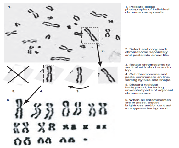
Figure 1: Karyotype Preparation Steps in the preparation of a karyotype from digital microphotographs of metaphase spread. Chinese hamster cells recloned from the Y-5 strain.