Characterization of a cell line is vital for determining its functionality and in proving its authenticity as pure cell line. Special attention must be paid to the possibility that the cell line has become cross-contaminated with an existing continuous cell line or misidentified because of mislabeling or confusion in handling DNA profiling. This has now become the major standard procedure for cell line identification, and a standard procedure with universal application.
The various important factors for cell line characterization are:
- It leads to authentication or confirmation that the cell line is not cross-contaminated or misidentified
- It is confirmation of the species of origin
- It is used for correlation with the tissue of origin, which comprises the following characteristics:
- Identification of the lineage to which the cell belongs
- Position of the cells within that lineage (i.e., the stem, precursor, or differentiated status)
- For determination whether the cell line is transformed or not:
- Whether the cell line is finite or continuous?
- Whether the cell line expresses properties associated with malignancy?
- It indicates whether the cell line is prone to genetic instability and phenotypic variation
- Identification of specific cell lines within a group from the same origin, selected cell strains, or hybrid cell lines, all of which require demonstration of features unique to that cell line or cell strain
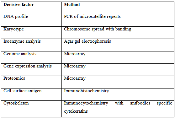
Table 1: Decisive factors for characterization of cell lines and corresponding methods
Parameters of Characterization
The nature of the technique used for characterization depends on the type of work being carried out. Some of the parameters are:
- In case molecular technology, DNA profiling or analysis of gene expression are most useful.
- A cytology laboratory may prefer to use chromosome analysis coupled with FISH (fluorescence in situ hybridization) and chromosome painting. Chromosomal analysis also known as karyotyping, is one of the best traditional methods for distinguishing among species. Chromosome banding patterns can be used to distinguish individual chromosomes. Chromosome painting, explicitly using combinations of specific molecular probes that hybridize to individual chromosomes, adds further resolution and specificity to this technique. These probes identify individual chromosome pairs and are species specific. Chromosome painting is a good method for distinguishing between human and mouse chromosomes in potential cross-contaminations.
- A laboratory with immunological capability may prefer to use MHC (Major Histo compatibility complex) analysis (e.g., HLA typing) coupled with lineage specific markers. Combined with a functional assay related to our own interests, these procedures should provide sufficient data to authenticate a cell line as well as confirm that it is suited to the concerned.
- Lineage or Tissue markers: The progression of cells down a particular differentiation pathway towards a specific differentiated cell type and can be considered as a lineage, and as cells progress down this path they acquire lineage markers specific to the lineage and distinct from markers expressed by the stem cells. These markers often reflect the embryological origin of the cells from a particular germ layer. Lineage markers are helpful in establishing the relationship of a particular cell line to its tissue of origin. There are some lineage markers which are described as follows:
- Cell surface antigen: These markers are particularly useful in sorting hematopoietic cells and have also been effective in discriminating epithelium from mesenchymally derived stroma with antibodies such as anti- and anti-HMFG 1 and, distinguishing among epithelial lineages, and identifying neuroectodermally derived cells (e.g., with anti-A2B5).
- Intermediate filament proteins : These are among the most widely used lineage or tissue markers. Glial fibrillary acidic protein (GFAP) for astrocytes and desmin for muscle are the most specific, whereas cytokeratin marks epithelial cells and mesothelium.
- Differentiated products and functions : Haemoglobin for erythroid cells, myosin or tropomyosin for muscle, melanin for melanocytes, and serum albumin for hepatocytes are examples of specific cell type markers, but like all differentiation markers, they depend on the complete expression of the differentiated phenotype.
Transport of inorganic ions, and the resultant transfer of water, is characteristic of absorptive and secretary epithelia. Polarized transport can also be demonstrated in epithelial and endothelial cells using Boyden chambers or filter well inserts. Other tissue-specific functions that can be expressed in vitro include muscle contraction and depolarization of nerve cell membrane.
- Enzymes: Three parameters are available in enzymatic characterization:
- The constitutive level (in the absence of inducers or repressors)
- The induced or adaptive level (the response to inducers and repressors)
- Isoenzyme polymorphisms
- Cell surface antigen: These markers are particularly useful in sorting hematopoietic cells and have also been effective in discriminating epithelium from mesenchymally derived stroma with antibodies such as anti- and anti-HMFG 1 and, distinguishing among epithelial lineages, and identifying neuroectodermally derived cells (e.g., with anti-A2B5).