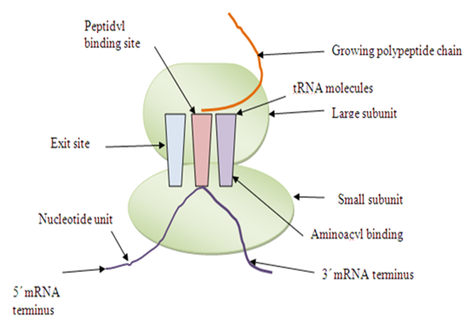
Figure 2: The detailed structure of a ribosome involved in protein synthesis. The figure is not upto the scale of ribosome.
Endoplasmic reticulum:
Endoplasmic reticulum is a network of interconnected internal membranes generally, the largest membrane in a eukaryotic cell—an extensive network of closed, flattened membrane-bounded sacs called cisternae (Figure 3). The name “endoplasmic reticulum” was coined in 1953 by Porter, who had observed it in electron micrographs of liver cells. The endoplasmic reticulum has a number of functions in the cell but is particularly important in the synthesis of lipids, membrane proteins, and secreted proteins.
Figure 3: The Endoplasmic reticulum.
Occurrence:
The occurrence of the endoplasmic reticulum is in eukaryotic cells with variation in its position from cell to cell. The erythrocytes (RBC), egg and embryonic cells lack in endoplasmic reticulum. ER is poorly developed in certain cells as the RBC which produces only proteins to be retained in the cytoplasmic matrix (haemoglobin), although the cell may contain many ribosomes). The spermatocytes also have poorly developed endoplasmic reticulum.
Morphology:
The endoplasmic reticulum occurs in three forms: 1. Lamellar form or cisternae which is a closed, fluid-filled sac, vesicle or cavity is called cisternae; 2. vesicular form or vesicle and 3. tubular form or tubules.
1. Cisternae: The cisternae are long, flattened, sac-like, unbranched tubules having diameter of 40 to 50 μm. They remain arranged parallely in bundles or stakes. RER mostly exists as cisternae which occur in those cells which have synthetic roles as the cells of pancreas, notochord and brain.
2. Vesicles: The vesicles are oval, membrane-bound vacuolar structures having diameter of 25 to 500 μm. They often remain isolated in the cytoplasm and occur in most cells but especially abundant in the SER.
3. Tubules: The tubules are branched structures forming the reticular system along with the cisternae and vesicles. They usually have the diameter from 50 to 190 μm and occur almost in all the cells. Tubular form of ER is often found in SER and is dynamic in nature, i.e., it is associated with membrane movements, fission and fusion between membranes of cytocavity network.
Ultrastructure:
The cavities of cisternae, vesicles and tubules of the endoplasmic reticulum are bounded by a
thin membrane of 50 to 60 Aº thickness. The membrane of endoplasmic reticulum is fluid-mosaic like the unit membrane of the plasma membrane, nucleus, Golgi apparatus. The membrane of endoplasmic reticulum remains continuous with the membranes of plasma membrane, nuclear membrane and Golgi apparatus. The cavity of the endoplasmic reticulum is well developed and acts as a passage for the secretory products. Palade in the year 1956 has observed secretory granules in the cavity of endoplasmic reticulum amking it rough in appearance. Sometimes, the cavity of RER is very narrow with two membranes closely apposed and is much distended in certain cells which are actively engaged in protein synthesis (acinar cells, plasma cells and goblet cells). The membranes of the endoplasmic reticulum contains many kinds of enzymes which are needed for various important synthetic activities. Some of the most common enzymes are found to have different transverse distribution in the ER membranes. The most important enzymes are the stearases, NADH-cytochrome C reductase, NADH diaphorase, glucose-6-phosphotase and Mg++ activated ATPase. Certain enzymes of the endoplasmic reticulum such as nucleotide diphosphate are involved in the biosynthesis of phospholipid, ascorbic acid, glucuronide, steroids and hexose metabolism.