1. Prophase:
The chromosomes are in the form of extended filaments and cannot be seen with a light microscope as discrete bodies except for the presence of one or more dark bodies (i.e. nucleoli) in the interphase stage. The beginning of prophase is marked by the condensation of chromosomes to form visibly distinct, thin threads within the nucleus. Each chromosome is already longitudinally double, consisting of two closely associated subunits called chromatids which are held together by centromere. Each pair of chromatids is the product of the duplication of one chromosome in the S period of interphase. As prophase progresses, the chromosomes become shorter and thicker as a result of intricate coiling. At the end of prophase, the nucleoli disappear and the nuclear envelope, a membrane surrounding the nucleus, abruptly disintegrates.
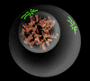
Figure 4: Prophase
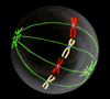
2. Metaphase:
At the beginning of metaphase, the mitotic spindle forms which are a bipolar structure and consist of fiber-like bundles of microtubules that extend through the cell between the poles of the spindle. Each chromosome attached to several spindle fibers in the region of the centromere. The structure associated with the centromere to which the spindle fibers attach is known as the kinetochore. After the chromosomes are attached to spindle fibers, they move towards the center of the cell until all the kinetochores lie on an imaginary plane equidistant from the spindle poles. This imaginary plane is called the metaphase plate. Hence the chromosomes reach their maximum contraction and are easiest to count and examine for differences in morphology. The signal for chromosome alignment comes from the kinetochore, and the chemical nature of the signal seems to be the dephosphorylation of certain kinetochore-associated proteins. The role of the kinetochore is demonstrated by the finding that metaphase is not delayed by an unattached chromosome whose kinetochore has been destroyed by a focused laser beam. The role of dephosphorylation is demonstrated through the use of an antibody that reacts specifically with some kinetochore proteins only when they are phosphorylated. Unattached kinetochores combine strongly with the antibody, but attachment to the spindle weakens the reaction. In chromosomes that have been surgically detached from the spindle, the antibody reaction with the kinetochore reappears. Through the signaling mechanism, when all of the kinetochores are under tension and aligned on the metaphase plate, the metaphase checkpoint is passed and the cell continues the process of division.
 |
 |
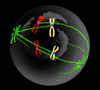
| Figure 5: Prometaphase |
Figure 6: Metaphase |