C. C-terminal residues: Not many methods are developed for c-terminal amino acid analysis. The most common method is to treat the protein with a carboxypeptidase to release the c-terminal amino acid and test the solution in a time dependent manner.
Stage 4. Ordering the peptide fragments: The usage of different protein cleavage reagent produces over-lapping amino acid stretches and these stretches can be used to put the whole sequence.
Stage 5. Locating disulfide bonds: The protein cleavage by typsin is performed with or without breaking di-sulphide linkage. Amino acid sequence analysis of the fragments will provide the site of disulphide bond. The presence of one disulphide will reduce two peptide fragment and will appear as one large peptide fragment.
Mass Spectrometry Method: In recent pass, mass spectroscopy in conjugation with proteomics information is also been popular tool to chacracterize each peptide fragment to deduce its amino acid sequence. The minor detail of this approach can be explored by following the article

Methods to determine secondary, tertiary structures:
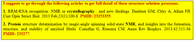
A. Experimental Methods: X-ray crystallography and NMR spectroscopy are the two methods can be used to determine the 3-dimensional structure of the target enzyme.
B. Homology modeling- This is a useful and fast structural solution method where the sequence similarities between the template and the target enzyme is used to model the 3-dimensional structure of the target enzyme. The homology modeling exploits the idea that the amino acid sequence of a protein directs the folding of the molecule to adopt a suitable 3-dimensional conformation with minimum free energy.