B. Labeling of Lysosome:
1. Plate cells on cover glasses in 24 well plate.
2. Grow them with 100ug rhodamine dextran O/N in DMEM + 10% FBS+1% antibiotics cocktails.
3. Wash the cells with PBS and chase for 1 Hrs in media without rhodamine dextran.
C. Fusion assay:
1. Add 10µg/ml 1 mM latex/IgG beads in 0.5ml media and spin at 1000G for 2 min.
2. Incubate for another 5 min in 370C in water bath.
3. Remove the beads and wash them two time with PBS at 370C.
4. Media is removed and fixed with 4% paraformaldehyde.
5. Slide were visualized in fluorescence microcope.
D. Observation: Observe the cells in the bright field and look for the beads on the cells. Observe the cells in the fluorescence microscope with UV filter. If the bead has blue fluorescence, then the cells can be visualized through red channel.
E. Analysis: A typical phagocytosis of bead will represent by the appearance of beads in the phase and the same bead will be circled by blue fluorescence from filipin (Figure 33.2). If the bead has blue fluorescence ring, and it further encircled by red ring indicates interaction of lysosome and phagosome (Figure 33.2).
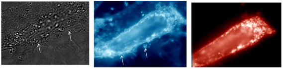
Figure 33.2 : Phagosome-lysosome interaction by fluorescence microscope. Arrow indicates the position of phagosome fused with lysosome.