5. Washing : The primary antibody needs to wash to reduce the background signal. Sample is washed with 2% BSA prepared in PBS.
6. Seconadry Staining: Incubate the sample with secondary antibody (1:500 in 2% BSA) for overnight at 40C or 1hrs at 370C.
7. Washing: The secondary antibody needs to wash to reduce the background signal. Sample is washed with 2% BSA prepared in PBS.
8. Mounting: The sample is sensitive to the loss of water and needs to preserve in a mounting media containing glycerol. In addition, fluorescence signal is sensitive to the high laser beam and it require protection by adding antifading agent. In a typical mounting media, glycerol containing PPD is used to mount fluorescent sample.
9. Observation and visualization: Sample is fixed on the microscope stage and then observe under bright light to check the cells morphology by turning focusing knob. If the sample’s morphology is good then it can observe under fluorescence channel (Figure 32.5).
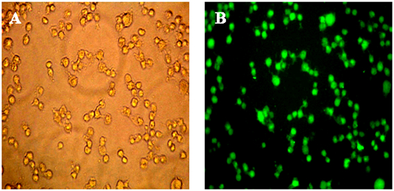
Figure 32.5: Bright-field (A) and epifluorescence (B) images of Cos-7 cells expressing GFP.