Protocol: Remove the cells from the cell culture plate by trypsinization or by 0.5% EDTA in PBS. Plate a small amount of cells on the glass slide and cover them with cover slip. Mix 50ml of cell suspension with the 50ml of trypan blue solution (0.4%) and fill the hemocytometer chamber. Observe the cells under the 20x objective using inverted microscope with phase plate. Trypan blue is a charged dye and viable cells exclude this dye to the presence of membrane potential where as dead cells (in the absence of membrane potential) accumulates the dye in the cytosol (Figure 32.3). Hence, viable cells appear colorless where as dead cells appear blue or dark colored.The hemocytometer is placed on the microscope stage and the cell suspension is counted. There is a "V" or notch at either end through which cell suspension is loaded into the hemocytometer. The cells are counted in the chambers and that gives the number of cells. In addition, blue colored cells can be counted to know the number of dead cells.
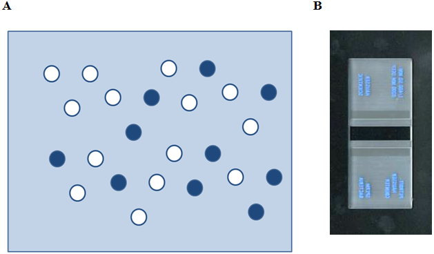
Figure 32.3 Observation of cell suspension after trypan blue staining. (A) Viable cells appears colorless where as dead cells takesup dye and appear dark blue. (B) Hemocytometer
Lab Experiment 32.1 : Immuno-localization
Background Information: Unlike the other types of light microscopy that need special optics to enhance the contrast, fluorescence in visible region of electromagnetic radiation is directly detected. The cellular features, however, can be studied using extrinsic fluorescent probes that can go inside the cell and bind to the intracellular molecules with high specificity. The fluorescence emission of the dyes used in biological microscopy span the entire visible region of the electromagnetic spectrum. Optical diagram of an epifluorescence microscope is given in the Figure 32.4. In an epifluorescence microscope, the illumination of the specimen as well as the collection of the fluorescence light is achieved by a single lens. This has become possible due to the incorporation of dichroic mirror in the optics. A dichroic mirror is largely reflective for the light below a threshold wavelength and transmissive for the light above that wavelength.
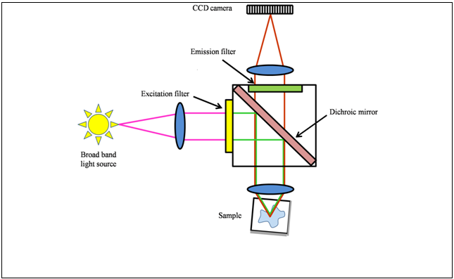
Figure 32.4: A diagram showing the optical path in an epifluorescence microscope.