
2. Native PAGE: SDS-PAGE discussed in the previous lecture is using anionic detergent sodium dodecyl sulfate and β-mercaptoethanol to give equal charge to all protein and breaks the disulphide linkage. As a result, the 3-D structure of the protein is destroyed and it migrate as per their subunit molecular weight. In the native PAGE, sample is prepared in the loading dye does not contains detergent or denaturating agent and as a result, sample runs on the basis of charge/mass. In native PAGE, the 3-D conformation as well as activity of the protein remains unaffected.
3. Urea PAGE: In this method, insoluable protein is dissolved in Urea and samples separate based on their charge/subunit mass. A gradient Ura PAGE is used to monitor the folding states of a protein.
2. Horizontal gel electrophoresis- The electrophoresis in this gel system is performed in a continuous fashion with both electrodes and gel cassette submersed within the buffer. The schematic diagram of a vertical gel electrophoresis apparatus is given in Figure 36.1. The electrophoresis chamber has two platinum electrodes placed on the both ends are connected to the external power supply from a power pack which supplies a direct current or DC voltage. The tank filled with the running buffer and the gel casted is submerged inside the buffer. There are additional accessories needed for casting the agarose gel such as comb (to prepare different well), spacer, gel caster etc.
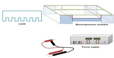
Figure 36.1: Different components of horizontal gel electrophoresis apparatus.