A secondary electron detector is biased with positive potential to attract the low energy secondary electrons. Detector for backscattered electrons is not biased; the high energy backscattered electrons strike the unbiased detector. As backscattered electrons come from a significant depth within the sample (Figure 18.3), they do not provide much information about the specimen topology. However, backscattered electrons can provide useful information about the composition of the sample; materials with higher atomic number produce brighter images.
Sample preparation for SEM: A specimen to be analyzed by electron microscopy has to be dry which most biological samples are not. As dehydration might lead to structural changes, the specimens are first fixed to preserve their structural features. Fixation is the first step and can be achieved using chemical methods such as fixation with glutaraldehyde or physical methods such as cryofixation in liquid nitrogen. The fixed specimens are then dehydrated usually by exposing them to an increasing gradient of ethanol (up to 100%). The specimens are then dried using critical point method. The dried specimens are then coated with a conducting material usually gold to make the surface conducting and cause it emit more secondary electrons. A SEM image of human erythrocytes coated with gold is shown in figure 18.4
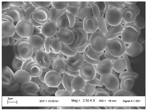
Figure 18.4 A scanning electron micrograph of human erythrocytes.