4. Protoplast development and regeneration
Protoplast starts to regenerate a cell wall within few days (2-4 days) of culture and during this process, protoplasts lose their characteristic spherical shape which has been taken as an indication of new wall regeneration. Cell wall regeneration can be confirmed by Calcofluor White staining method. There is direct relationship between wall formation and cell division. Protoplasts which are not able to regenerate a proper wall fail to undergo normal mitosis. Protoplasts with a poorly developed wall often show budding and may enlarge several times their original volume. They may become multinucleate because karyokinesis is not accompanied by cytokinesis. Among other reasons, inadequate washing of the protoplasts prior to culture leads to these abnormalities.
And completes process when provided with suitable condition of light, pH and temperature newly synthesized protoplast can be visualized by staining. Once the cell wall formation is completed, cells undergo division resulting in increase size of cells. After an interval of 3 weeks, small cell colonies appear, these colonies are transferred to an osmotic-free callus induction medium. This is followed by introduction into organogenic or embryogenic medium leading to plantlet development.
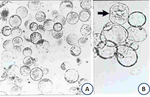
Figure 12.4: Protoplast isolation and cell wall regeneration. A. Isolated protoplast showing spherical structure; B. Wall is regenerated around the protoplast and one of the protoplasts showing cell division (arrow marked)