Procedure :
1. Prepare a smear on a microscopic slide from the clinical sample.
2. Keep the slide on the staining rack.
3. Flood the slide with strong Carbolfuchsin for 5-7 minutes with intermittent heating until steam rises. Avoid boiling or drying of the stain (Fig. 14)
4. Wash with water.
5. Decolorize with 20% H2SO4 for 2 minutes.
6. Wash with water.
7. Add counter stain methylene blue for 30 seconds
8. Acid-fast bacteria appear red, and non-acid-fast bacteria (and other organisms and cellular materials) appear blue (Fig. 15).
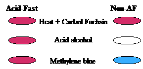
Fig. 14. Acid fast staining procedure
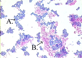
Fig. 15. A. Non acid fast bacteria B. Acid fast bacteria
Special stains
Special stains use a variety of dyes and techniques to stain particular tissues, structures or pathogens (such as bacteria) to assist pathologists with tissue-based diagnosis. Special stains can be applied to cell biology and histology. e.g. Acid fuchsin, Congo red and Malachite green.
Some useful applications are : The determination of DNA and RNA content, the mode of action of drugs, hormones or of potentially toxic food additives, metabolic biochemistry, and biochemistry of disease Processes, identification of micro-organisms.
Staining specific structures:
Spore staining:
• Bacteria in the genera Bacillus and Clostridium have dormant structures called endospores.
• Endospore morphology and location vary with species and often are valuable in identification.
• Most commonly used method for endospore stain is the Schaeffer-Fulton endospore stain method.
• Malachite green, the primary stain is applied to a heat-fixed smear and heated to steaming for about 5 min (heat helps the stain to penetrate the endospore wall).
• Preparation is washed off with water to remove malachite green from all of the cells parts except the endospores.
• Next, safranin is applied to the smear to stain portions of the cell other than endospores.
• The endospores appear green within red or pink cells (Fig. 16).
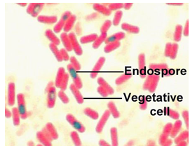
Fig. 16. Endospore staining