Gram staining
Differential staining requires the use of chemical reagents that are applied sequentially to a heat-fixed smear. The first reagent is called the primary stain. Its function is to impart color to all cells. In order to establish a color contrast, the second reagent used is the decolorizing agent. Based on the chemical composition of the cellular components, the decolorizing agent may or may not remove the primary stain from the entire cell or only from certain cell structures. The final reagent, counter stain , has a contrasting color to that of the primary stain. Following decolorization, if the primary stain is not washed out, the counter stain cannot be absorbed and the cell or its components will retain the color of the primary stain. If the primary stain is removed, the decolorized cellular components will accept and assume the contrasting color of the counterstain. The most important differential stain used in bacteriology is the Gram stain. It divides bacterial cells into two major groups, gram-positive and gram-negative, which makes it an essential tool for classification and differentiation of microorganisms.
Primary stain
Crystal Violet
This is used first and stains all cells purple.
Mordant
Gram's Iodine
This serves as a substance that forms an insoluble complex by binding to primary stain. The resultant crystal violet-iodine (CV-I) complex serves to intensify the color of the stain, and all the cells will appear purple-black at this point.
Decolorizing Agent
Ethyl alcohol, 95%
The reagent serves a dual function as a lipid solvent and as a protein dehydrating agent. Its action is determined by the lipid concentration of the microbial cell wall. In gram-positive cells, the low lipid concentration is important for retention of the CV-I complex. Therefore, the small amount of lipid content is readily dissolved by the action of the alcohol, forming minute cell wall pores. These are then closed by alcohol's dehydrating effect. Consequently, the tightly bound primary stain is difficult to remove, and the cells remain purple. In gram-negative cells, the high lipid concentration found in outer layers of the cell wall is dissolved by the alcohol, creating large pores in the cell wall that do not close appreciably on dehydration of cell wall proteins. This facilitates release of the unbound CV-I complex, leaving those cells colorless or unstained.
Counterstain
Safranin
This is the final reagent, used to stain red those cells that have previously decolorized. Since only gram-negative cells undergo decolorization, they may now absorb the counterstain. Gram-positive cells retain the purple color of the primary stain.
PROCEDURE
- One or two loopful of suspended cells was applied directly to the glass slide with a sterile inoculating loop and spread evenly (Fig. 12).
- The smear was then heat fixed by rapid passage of the air-dried smear two-three times over the flame of the Bunsen burner.
- The heat fixed smear was flooded with crystal violet and incubated for 1 minute.
- Crystal violet was washed away with distilled water.
- The smear was flooded with Gram's iodine mordant and incubated for 1 minute.
- The mordant was washed away with distilled water.
- Ethyl alcohol (95%) was added drop wise to the smear until crystal violet fails to wash off.
- The alcohol was washed away with distilled water.
- The smear was counterstained with safranin for 45 seconds.
- The counterstain was washed away with distilled water.
- The slide was dried (either blot dried or in an incubator) and examined in a compound microscope (Fig. 13).
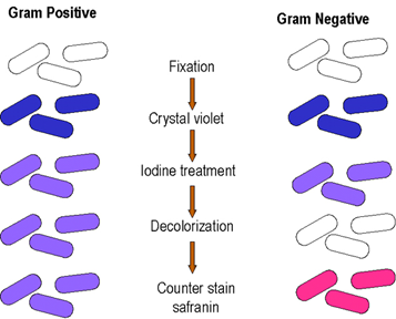
Fig. 12 . Procedure of Gram staining
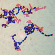
Fig. 13 . Gram positive and negative cells when viewed under microscope
Acid fast staining techniques principle :
Most of the bacteria can be stained with aqueous solutions of para rosaniline dyes but certain organisms especially which belongs to family mycobacterium resist entry of these weaker dyes and hence these weak solutions are unsuitable for staining them. They can be stained with strong solutions which contain phenols which help in penetration of the in to the cell. Heating further enhances entry of the dye in to the cell. These organisms once stained resist decolorization with acids such as H2SO4, HCL, HNO3 etc. and even with alcohol. Those organisms which resist decolorize with acid retain primary stain and are called acid fast organisms and those which are decolorized by acid will take up counter stain and are called non acid fast organisms. Reason for acid fastness is due to the presence of long chain fatty acid, mycolic acid in the cell wall of these organisms.