Structure:
Transmission electron microscopic studies have shown that the bacterial flagellum is composed of three parts. 1) Filament – outermost region and contain the globular protein flagellin2) Hook – the filament is attached to hook, which consists of a different protein 3) Basal body - which anchors the flagellum to the cell wall and plasma membrane. It consists of a small central rod inserted into it are a series of rings (Fig. 16). The filament is a hollow, rigid cylinder constructed of a single protein called flagellin (MW from 30,000 to 60,000). Some bacteria have sheaths surrounding their flagella. For example Bdellovibrio has a membranous structure surrounding the filament. Vibrio cholerae has a lipopolysaccharide sheath.
The hook and basal body are quite different from the filament. Slightly wider than the filament, the hook is made of different protein subunits. The basal body is the most complex structure of the flagellum. In E.coli and Gram negative bacteria, the body has four rings connected to central rod. The outer L and P rings associate with the lipopolysaccharide and peptidoglycan layers. The inner M ring connects the plasma membrane.Gram positive have only twp basal body rings, an inner ring connected to the plasma membrane and an outer one probably attached to the peptidoglycan.
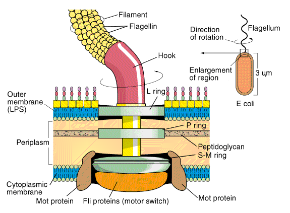
Fig. 16. Structure of bacterial flagella (Gram negative)
The synthesis of flagella is a complex process involving atleast 20 to 30 genes. Flagellin subunits are transported through the filament's hollow internal core. When they reach the tip, the subunits spontaneously aggregate under the direction of a special filament cap so that the filament grows at its tip rather than at the base. Filament synthesis is an excellent example of self-assembly.
Flagellar movement:
The mechanism of flagellar movement in prokaryotes is different from eukaryotic flagella. The bacterium moves when the helix rotates as the filament is in the shape of rigid helix. The flagella act just like propellers on a boat. The direction of flagellar rotation determines the nature of bacterial movement. The movement in monotrichous bacteria stop and tumble randomly by reversing the flagellar rotation. The polar flagella, rotate counter clockwise during normal forward movement, whereas the cell itself rotates slowly clockwise. Peritrichous bacteria also operate in a similar way. To move forward, the flagella rotate counter clockwise. As they do so, they bend at their hooks to for a rotating bundle that propels them forward. Clockwise rotation of the flagella disrupts the bundle and the cell tumbles (Fig. 17).
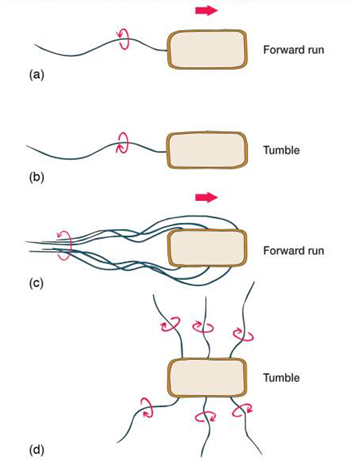
Fig. 17. Bacterial flagellar movement