In this lecture we shall look into the structures external to the cell wall of bacterial cells. This includes glycocalyx, fimbriae, pili, flagella, axial filaments
Glycocalyx (Capsules, Slime layers and S-layers)
It is a viscous (sticky), gelatinous polymer composed of polysaccharide, polypeptide or both. If the substance is organized and is firmly attached to the cell wall, the glycocalyx is described as a capsule (negative staining). If the substance is unorganized and only loosely attached to the cell wall, the glycocalyx is described as a slime layer. Capsules protect pathogenic bacteria from phagocytosis (process by which certain white blood cells engulf and destroy microbes) and contribute to virulence. Unencapsulated Streptomyces pneumoniae and Bacillus anthracis does not cause disease because the cells are readily phagocytosized. This allows the bacteria to attach to various surfaces, such as rocks in fast-moving streams, plant roots, human tooth and tissues and even other bacteria. Capsules also contain water which prevents them from desiccation. Other examples are Streptococcus mutans (dental caries), Klebsiella pneumoniae (respiratory tract). These can protect a cell against dehydration. Capsules and slime layers usually are made up of polysaccharides, but they may be constructed of othermaterial, like Bacillus anthracis has a capsule of poly D-glutamic acid. Capsules are clearly visible in the light microscope by using stains or special capsule stains.
A regularly structured layer called S-layer is usually seen in many gram positive and gram negative bacteria. It consists of proteins or glycoproteins and resembles a pattern something similar to floor tiles. The S-layer adheres directly to the outer membrane in case of gram negative bacteria and with the peptidoglycan surface in gram positive bacteria. These protect the bacteria against ion and pH fluctuations, osmotic stress, enzymes, or the predacious bacterium Bdellovibrio . The S layer also helps maintain the shape and envelope rigidity of at least bacterial cells and also promotes cell adhesion to surfaces. Sometimes, the layer also seems to protect some pathogens against complement attack and phagocytosis, thus contributing to their virulence.
Fimbriae and Pili:
Many gram negative bacteria have hairlike appendages that are shorter, straighter and thinner than flagella and are used for attachment rather than for motility. They are usually called fimbriae. These structures contain a protein called pilin. Fimbriae - occur at the poles of the bacterial cell, or they can be evenly distributed over the entire surface of the cell. Fimbriae of Neisseria gonorrhoeae the causative agent of gonorrhea help the microbe to colonize mucous membranes to cause the disease. At least some types of fimbriae attach bacteria to solid surfaces such as rocks in streams and host tissues.
Pilior sex pili or pilus - usually longer than fimbriae and number only one to ten per cell. Pili function to join bacterial cells prior to the transfer to DNA from one cell to another (sometimes called sex pili). They are genetically determined by sex factors or conjugative plasmids and are required for bacterial mating. Some bacterial viruses attach specifically to receptors on sex pili at the start of their reproductive cycle.
Flagella:
Motile bacteria move by use of flagella, threadlike locomotor appendages extending outward from the plasma membrane and cell wall. They are slender, rigid structures, about 20 nm across and up to 15 or 20 µm long. Bacterial species often differ distinctively in their patterns of flagella distribution (Fig. 15).
Monotrichous- single polar flagellum located at one end
Amphitrochous- With two flagella, one at each end
Lophotrichous - With two or more flagella at one or both ends
Peritrichous - flagella all over the surface
Atrichous - Bacteria without flagella (Cocci rarely have flagella)
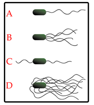
Fig. 15 . Flagellar arrangement. A. Monotrichous B. Lophotrichous C. Amphitrichous D. Peritrichous