Prokaryotic cells
Prokaryote means before nucleus in Greek. They include all cells which lack nucleus and other membrane bound organelles. Mycoplasma, virus, bacteria and cyanobacteria or blue-green algae are prokaryotes.
Most prokaryotes range between 1 μm to 10 μm, but they can vary in size from 0.2 μm to 750 μm (Thiomargarita namibiensis). They belong to two taxonomic domains which are the bacteria and the archaea. Most prokaryotes are unicellular, exceptions being myxobacteria which have multicellular stages in their life cycles. They are membrane bound mostly unicellular organisms lacking any internal membrane bound organelles. A typical prokaryotic cell is schematically illustrated in Figure 1. Though prokaryotes lack cell organelles they harbor few internal structures, such as the cytoskeletons, ribosomes, which translate mRNA to proteins. Membranous organelles are known in some groups of prokaryotes, such as vacuoles or membrane systems devoted to special metabolic properties, e.g., photosynthesis or chemolithotrophy. In addition, some species also contain protein-enclosed microcompartments, which have distinct physiological roles (carboxysomes or gas vacuoles).
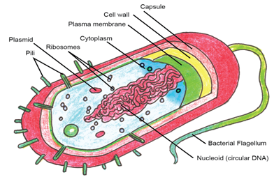
Figure 1: Schematic diagram of a prokaryotic cell
The individual structures depicted in Figure 1 are as follows and details will be discussed in forthcoming chapters:
Flagella: It is a long, whip-like protrusion found in most prokaryotes that aids in cellular locomotion. Besides its main function of locomotion it also often functions as a sensory organelle, being sensitive to chemicals and temperatures outside the cell.
Capsule: The capsule is found in some bacterial cells, this additional outer covering protects the cell when it is engulfed by phagocytes and by viruses, assists in retaining moisture, and helps the cell adhere to surfaces and nutrients. The capsule is found most commonly among Gram-negative bacteria. Escherichia coli, Klebsiella pneumoniae Haemophilus influenzae, Pseudomonas aeruginosa and Salmonella are some examples Gram-negative bacteria possessing capsules. Whereas examples of Gram positive bacteria are Bacillus megaterium, Streptococcus pneumoniae, Streptococcus pyogene.
Cell wall: Cell wall is the outermost layer of most cells that protects the bacterial cell and gives it shape. One exception is Mycoplasma which lacks cell wall. Bacterial cell walls are made of peptidoglycan which is made from polysaccharide chains cross-linked by unusual peptides containing D-amino acids. Bacterial cell walls are different from the cell walls of plants and fungi which are made of cellulose and chitin, respectively. The cell wall of bacteria is also distinct from that of Archaea, which do not contain peptidoglycan. The cell wall is essential to the survival of many bacteria. The antibiotic penicillin is able to kill bacteria by preventing the cross-linking of peptidoglycan and this causes the cell wall to weaken and lyse. Lysozyme enzyme can also damage bacterial cell walls.
There are broadly speaking two different types of cell wall in bacteria, called Gram-positive and Gram-negative (Figure 2). The names originate from the reaction of cells to the Gram stain, a test long-employed for the classification of bacterial species. Gram-positive bacteria possess a thick cell wall containing many layers of peptidoglycan and teichoic acids. In contrast, Gram-negative bacteria have a relatively thin cell wall consisting of a few layers of peptidoglycan surrounded by a second lipid membrane containing lipopolysaccharides and lipoproteins. These differences in structure can produce differences in property as antibiotic susceptibility. For example vancomycin can kill only Gram-positive bacteria and is ineffective against Gram-negative pathogens, such as Pseudomonas aeruginosa or Haemophilus influenzae.