Evidence of Stacking interactions: Compounds that interfere with Hydrogen bonds (urea, formamide) don’t separate strands by themselves, still requires heat.
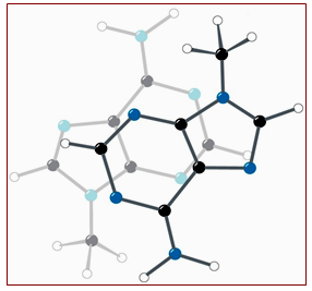
Figure 4.12: The Stacking of adenine rings in the crystal structure of 9-methyladenine.
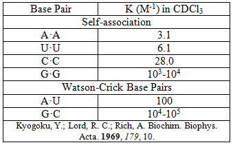
Table 4.1: Association Constants for Base Pair Formation.
- Charge-Charge Interactions- refers to the electrostatic (ion-ion) repulsion of the negatively charged phosphate is potentially unstable, however the presence of Mg2+ and cationic proteins with abundant Arginine and Lysine residues that stabilizes the double helix. Double-stranded helix structure thus, promoted by having phosphates on outside, interact with H2O and counter ions (K+, Mg2+, etc.).
- Solvation also plays a role in stabilizing the double helix that affects base pairing to mediating binding events.
Thus, the DNA strands in a double helix are held together by the H-bonds between the bases. The H-bonds already provide specificity but they also confer stability to the structure. The phosphate groups must be neutralized (by Na+ or Mg2+ ions) to allow the negatively charged phosphates to be in close proximity. The hydrophobic interactions between the planar base pairs stabilize the bases on the inside of the helix, so these provide stability to the structure but do not contribute to the specificity.
State-of-the-art experimental techniques, including denaturation study, study of salt effect are used to explore the binding affinities of DNA double-stranded oligomers in the solution. Besides these, electrospray ionization, liquid chromatography mass spectrometry (LCMS) and Fourier transform mass spectrometry (FTMS), are used to explore the binding affinities of DNA double-stranded oligomers in the gas phase, in the absence of solvent.
DNA Denaturation
DNA "Melting" is the term given to the separation of the two DNA strands. There are a number of ways to do this experimentally: Helical formation can be monitored by observing the optical density of a solution. The disruption of base stacking alters the electronic interaction between the bases. As the electronic interaction decreases, it becomes easier for an electron to absorb a photon. Hence, denaturation of DNA leads to the “hyperchromic” effect, i.e., the increased absorption of light. Thus, an increase in Temperature - when temperature of a DNA solution increases to the melting point (Tm), the strands separate. The absorbance of the solution changes as shown in the graph below. The increase in absorbance as the strands separate is due to the irregular orientation of the bases in the SS-DNA compared to the regular planar orientation in the helix.
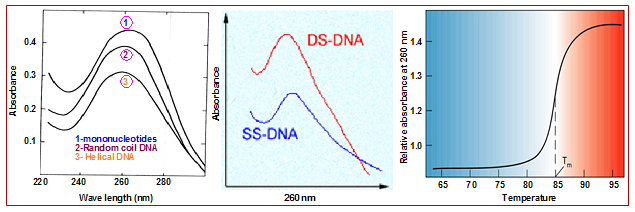
Figure 4.13: Temperature dependent absorbance and the melting temperature of DNA.