Electron Microscopy-I
Lab experiment 34.1: Preparation and analysis of the E.coli bacterium cells in scanning electron microscope.
Background Information: There are two basic models of the electron microscopes: Scanning electron microscopes (SEM) and transmission electron microscopes (TEM). In a SEM, the secondary electrons produced by the specimen are detected to generate an image that contains topological features of the specimen. The image in a TEM, on the other hand, is generated by the electrons that have transmitted through a thin specimen. Let us see how these two microscopes work and what kind of information they can provide:
Scanning electron microscope: Figure 34.1 shows a simplified schematic diagram of a SEM. The electrons produced by the electron gun are guided and focused by the magnetic lenses on the specimen.
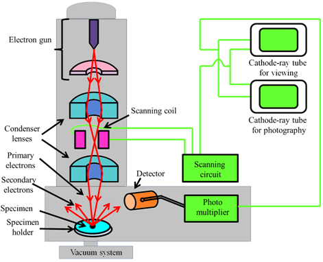
Figure 34.1 A simplified schematic diagram of a scanning electron microscope. |
The focused beam of electrons is then scanned across the surface in a raster fashion. This scanning is achieved by moving the electron beam across the specimen surface by using deflection/scanning coils. The number of secondary electrons produced by the specimen at each scanned point are plotted to give a two dimensional image. In principle, any of the signals generated at the specimen surface can be detected. Most electron microscopes have the detectors for the secondary electrons and the backscattered electrons. As backscattered electrons come from a significant depth within the sample, they do not provide much information about the specimen topology. However, backscattered electrons can provide useful information about the composition of the sample; materials with higher atomic number produce brighter images.