Background Information: The complex biological samples are efficiently resolved in 2-D gel electrophoresis. It involves the combination of charge and molecular weight to provide much greater separation in comparison to use of the individual property. The 2-D electrophoresis is a combination of isoelectrofocusing (IEF) followed by SDS-PAGE in a perpendicular direction ( Figure 16.1 ). The isoelectric point (pI) seperates the sample based on their isoelectric pH (it is indirecelty related to the charge present on the protein) where as SDS-PAGE seperates the molecules based on sizse (it is indirectly related to the molecular weight). In general analysis of complex bacterial lysate or tissue extract produces 1000-2500 well separated spots. With a sensitive detection tool and image analysis software, individual can be identified and compared under different conditions.
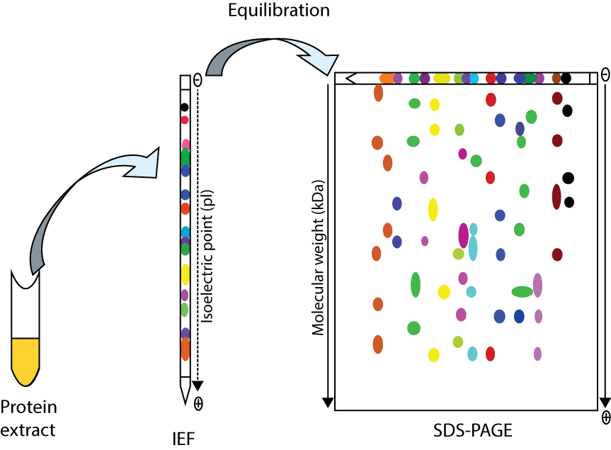
Figure 16.1: An Over-view of 2-Dimensional Gel Electrophoresis. needs redraw from
Lab Experiment 16.1 : Analyze the complex E.coli lysate in 2-D Gel electrophoresis.
Material and methods:
IPG
Reagents for SDS-PAGE
Reagents for silver stain
Trypsin
MALDI-Tof