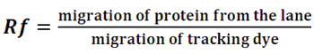
Running of the gel: The sample is prepared in the loading dye containing SDS, β-mercaptoethanol in glycerol to denature the sample and presence of glycerol facilitates the loading of sample in the well. As the samples are filled vertically there is a distance drift between the molecules at the top Vs at the bottom in a lane. This problem is taken care once the sample run through the stacking gel. The pH of the stacking gel is 6.8 and at this pH, glycine is moving slowly in the front where as Tris-HCl is moving fast. As a result, the sample gets sandwiched between glycine-Tris and get stacked in the form of thin band. As the sample enters into the resolving gel with a pH 8.8, the glycine is now charged, it moves fast and now sample runs as per their molecular weight (due to SDS they have equal negative charge). After tracking dye reaches to the bottom of the gel, gel is taken out from the glass plate with the help of a spatula. Gel is stained with coomassie brilliant blue R250 dye. The dye stains protein present on the gel. A typical SDS-PAGE pettern is given in the Figure 13.6 .
Potentials of discontinuous PAGE :
1. Number of disulfide bonds: Comparison of reducing and non-reducing denaturing gels can be used to provide information related to the number of disulfide bonds present in the protein.
2. Seperating Proteins based on size alone: In the presence of SDS and reducing environment, PAGE gel resolve two proteins of on the basis of molecular masses and the concentration of gel concentration.
In SDS-PAGE, the relative mobility and the log molecular weight as given by
|
|
(13.5) |
Molecular weight of a protein can be determined by plotting relative migration Rf with the log molecular weight of standard protein.
 |
(13.6) |
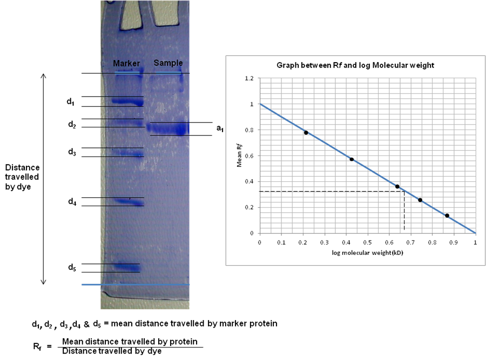
Figure 13.6: Determination of molecular weight using SDS-PAGE. (A) SDS-PAGE (B) Determinaion of Rf.