·
31.3 Dystrophin protein
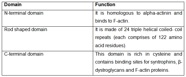
Dystrophin (a rod-shaped cytoplasmic protein) is a key player that connects the cytoskeleton of each muscle fiber to the basal lamina, with the help of a protein complex (made of numerous protein subunits more than 50), and thus provides mechanical stability by anchoring and supporting the sarcolemma. The dystrophin protein is made of three domains with different functionalities.
The dystrophin glycoprotein complex (DGC) also called costamere, acts as a bridge between the cytoskeleton, plasma membrane and the basal lamina. The dystroglycan is made of two subunits (α, β). The α-dystroglycan binds to laminin-2 (merosin) protein whereas the β-dystroglycan binds to the cysteine rich region of dystrophin. Sarcoglycans bind to the biglycans and the collagen and forms a tight complex. Syntrophins (peripheral membrane protein) bind to the C-terminal region of the dystrophin. The structure of DGC has been illustrated in the figure 31.1.
The absence of dystrophin results in excess calcium permeation to the sarcolemma (cell membrane). Due to the aberration in these signaling pathways, water enters into the mitochondria causing it to burst. This results in mitochondrial dysfunction causing an increase in stress-induced cytosolic calcium signals. As a result of amplification of stress-induced cytosolic calcium signals the production of stress-induced reactive-oxygen species (ROS) takes place. Following the ROS formation, the cascade activation of various complex pathways (not clearly understood) takes place and there is an upregulation of oxidative stress within the cell that causes damage to the sarcolemma and cell death. A large number of cells die as a result muscle fibre necrosis can be observed in biopsy. The dead muscles are replaced with adipose and connective tissue.