·
40.2 Labeling of cells
There are certain antigens specific for particular cell types. Antibodies against those antigens are used to identify the cells and tissues from a mixed population. The antibodies are labeled with fluorochrome, radioisotopes, or enzymes in order for their detection through different sensitive detector system.
40.2.1 Flow Cytometry
The cell lineages, stages of maturation, and types can often be determined by expression of molecules over the cell surface. The method generally involves staining the cells with fluorescence dye specific against the expressed molecules over the cell surface. The surface molecules are often termed as markers or “ cluster of differentiation ” ( CD ). The flow cytometer is a specific instrument which can detect the number of cells expressing the molecules based on the fluorescence detected by sensors. Occasionally cytoplasmic molecules are stained by fluorochrome- labeled antibody after permeabilizing the cells which permits the entry of labeled antibodies. DNA can also be labeled with propidium iodide to study the cell cycle. The dying cells can be labeled with annexin-V which binds with phosphatidylserine expressed in the cells undergoing apoptosis. It is a powerful tool to analyze the cell types and the cells which express different epitopes.
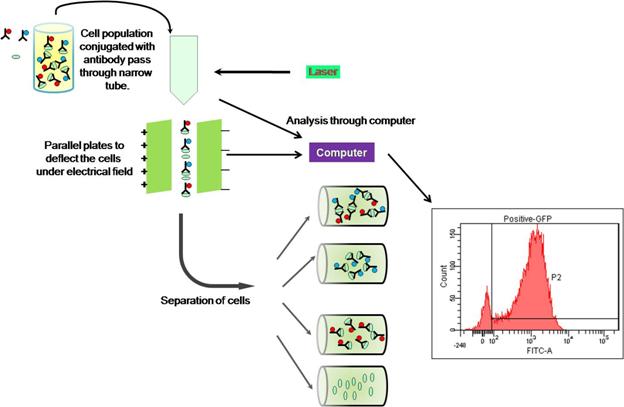
A more advanced technique called fluorescent- activated cell sorter is used to separate different cell types based on the intensity of the fluorescence on electro-magnetic field.
Figure 39.1 Schematic representation of fluorescent- activated cell sorter assay: