·
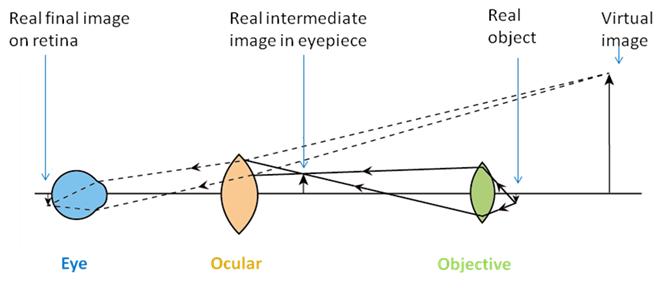
Figure 17.2: Perception of a magnified virtual image of a specimen in a microscope
The objective lens forms a magnified image of the object (called the real intermediate image) in or near the eye piece; the intermediate image is examined by the eyepiece and the eye, which forms a real image on the retina.
1.1. Resolving power of microscope
Resolution or resolving power of a microscope can be defined as the smallest distance apart at which two points on a specimen can still be seen separately. Sometimes, blurred images may be seen through the lenses. This is because of the placing of two distinct points too close to each other, which results in overlapping of the images. Magnification can neither improve nor decrease the resolving power of the microscope.
The resolution R is determined essentially by three parameters:
i. the wavelength λ of the illuminating light
ii. the numerical aperture (NA) of the objective lens (NAobj)
iii. the numerical aperture of the condenser (NAcond)
where,
R = 1.22*λ / (NAobj + NAcond) (i)
When the aperture of the condenser is adjusted to that of the objective, i.e. the aperture of the condenser is essentially same as the objective aperture, the equation (i) simplifies to:
R = 0.61*λ/NAobj (ii)
The resolution of an image is limited by the wavelength of radiation used to view the sample. This is because when objects in the specimen are much smaller than the wavelength of the radiation being used, they do not interrupt the waves, and so are not detected. The wavelength of light is much larger than the wavelength of electrons, so the resolution of the light microscope is a lot lower. Using a microscope with a more powerful magnification will not increase this resolution any further. It will increase the size of the image, but objects closer than 200nm will still only be seen as one point.