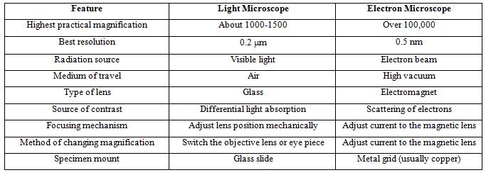
Table 1. Features and differences between light and electron microscope
Scanning Electron Microscope (SEM)
The Scanning Electron Microscope (SEM) produces images by detecting secondary electrons which are emitted from the surface due to excitation by the primary electron beam. In the SEM, the electron beam is scanned across the surface of the sample in a raster pattern, with detectors building up an image by mapping the detected signals with beam position (Fig. 9). The SEM image relies on electron interactions at the surface rather than transmission it is able to image bulk samples and has a much greater depth of view, and so can produce images that are a good representation of the 3D structure of the sample. SEM images are therefore considered to provide us with 3D, topographical information about the sample surface but will still always be only in black and white.This is specially used to study the external surface features of microorganisms.
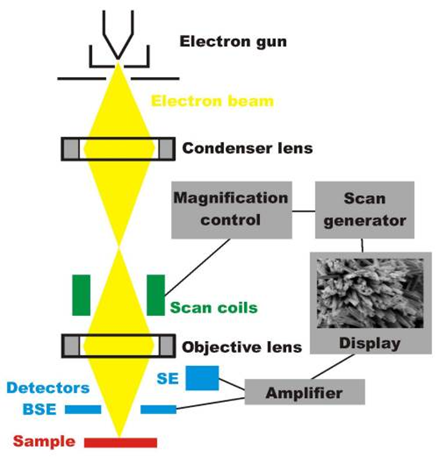
Fig. 9. Scanning electron microscope
Disadvantages of Electron Microscopy
Electron microscopes are very expensive to buy and maintain. They are dynamic rather than static in their operation: requiring extremely stable high voltage supplies, extremely stable currents to each electromagnetic coil/lens, continuously-pumped high/ultra-high vacuum systems and a cooling water supply circulation through the lenses and pumps. As they are very sensitive to vibration and external magnetic fields, microscopes aimed at achieving high resolutions must be housed in buildings with special services.
New microscopy technique
Confocal Microscopy
Confocal microscopy is an optical imaging technique used to increase optical resolution and of a micrograph by using point illumination and a spatial pinhole to eliminate out-of-focus light in specimens that are thicker than the focal plane. It enables the reconstruction of three-dimensional structures from the obtained images. This technique has gained popularity in the scientific and industrial communities and typical applications are in life sciences, semiconductor inspection and materials science
Three types of confocal microscopes are commercially available.
Confocal laser scanning microscopes use a pair of mirrors (one for the x and the other for the y axis) to scan the laser across the sample and "descan" the image across a fixed pinhole and detector.
• Spinning-disk ( Nipkow disk ) confocal microscopes use a series of moving pinholes on a disc to scan spot of light.
• Programmable Array Microscopes (PAM) uses an electronically controlled spatial light modulator (SLM) that produces a set of moving pinholes. The SLM is a device containing an array of pixels with some property (opacity, reflectivity or optical rotation) of the individual pixels that can be adjusted electronically. The SLM contains micro-electromechanical mirrors or liquid crystal components. The image is usually acquired by a CCD camera.
Each of these classes of confocal microscope has particular advantages and disadvantages. Most systems are either optimized for either recording speed (i.e. video capture) or high spatial resolution. Confocal laser scanning microscopes can have a programmable sampling density and very high resolutions while Nipkow and PAM use a fixed sampling density defined by the camera's resolution. Imaging frame rates are typically very slow for laser scanning systems (e.g. less than 3 frames/second). Commercial spinning-disk confocal microscopes achieve frame rates of over 50 per second – a desirable feature for dynamic observations such as live cell imaging. In practice, Nipkow and PAM allow multiple pinholes scanning the same are in parallel as long as the pinholes are sufficiently far apart. Cutting-edge development of confocal laser scanning microscopy now allows better than standard video rate (60 frames/second) imaging by using multiple micro-electromechanical systems -based scanning mirrors. Confocal X-ray fluorescence imaging is a newer technique that allows control over depth, in addition to horizontal and vertical aiming, for example, when analyzing buried layers in a painting