Monitoring For Contamination:
Potential sources of contamination are enumerated along with the precautions that should be taken to avoid them. Even in the best laboratories contaminations do arise, so the following procedure is generally recommended:
- Contamination by eye and with a microscope at each handling of a culture should be checked properly.
- If it is suspected that a culture is contaminated and the fact cannot be confirmed in situ, the hood or bench should be kept clear except suspected culture and Pasteur pipettes. Because of the potential risk to other cultures, this should be better to do after all your other culture work is finished. A sample should be removed from the culture and placed on a microscope slide. Slide should be checked with a microscope, preferably by phase contrast. If it is confirmed that the culture is contaminated, pipettes should be discarded, hood or bench should be swabbed with 70% alcohol containing a phenolic disinfectant. The hood or bench should not be used until the next day.
- Nature of the contamination should be recorded.
- If the contamination is new and is not widespread, the culture, the medium bottle used to feed it, and any other reagent (e.g., trypsin) that has been used in conjunction with the culture should be discarded properly into disinfectant, preferably in a fume hood and outside the tissue culture area.
- If the contamination is new and widespread all media, stock solutions, trypsin, and so forth in current use should be discarded immediately.
- If the same kind of contamination has occurred before check stock solutions for contamination (a) by incubation alone or in nutrient broth (b) by plating out the solution on nutrient agar. If (a) and (b) prove negative, but contamination is still suspected, 100 mL of solution should be incubated, filtered it through a 0.2-μm filter, and plated out filter on nutrient agar with an uninoculated control.
- If the contamination is widespread, multispecific, and repeated then one should check (a) the laboratory's sterilization procedures (e.g., the temperatures of ovens and autoclaves, particularly in the center of the load, the duration of the sterilization cycle), (b) the packaging and storage practices, (e.g., unsealed glassware should be resterilized every 24 h), and (c) the integrity of the aseptic room and laminar-flow hood filters.
- One should not be attempting to decontaminate cultures unless they are irreplaceable.
Visible Microbial Contamination: Characteristic features of microbial contamination are as follows:
- A sudden change in pH, usually a decrease with most bacterial infections, very little change with yeast until the contamination is heavy, and sometimes an increase in pH with fungal contamination.
- Cloudiness in the medium, sometimes with a slight film or scum on the surface or spots on the growth surface that dissipate when the flask is moved
- Under a 10X objective, spaces between cells will appear granular and may shimmer with bacterial contamination. Yeasts appear as separate round or ovoid particles that may bud off smaller particles. Fungi produce thin filamentous mycelia and, sometimes, denser clumps of spores which may be blue or green. With toxic infection, some deterioration of the cells will be apparent.
- Under a 100X objective, it may be possible to resolve individual bacteria and distinguish between rods and cocci. At this magnification, the shimmering that is visible in some infections will be seen to be caused by mobility of bacteria. Some bacteria form clumps or associate with the cultured cells.
- With a slide preparation, the morphology of the bacteria can be resolved with a 100× objective, but this is not usually necessary. Microbial infection may be confused with precipitates of media constituents (particularly protein) or with cell debris, but can be distinguished by their regular morphology. Precipitates may be crystalline or globular and irregular and are not usually as uniform in size. Clumps of bacteria may be confused with precipitated protein, but, particularly if shaken, many single or strings of bacteria will be seen. If you are in doubt, plate out a sample of medium on nutrient agar.
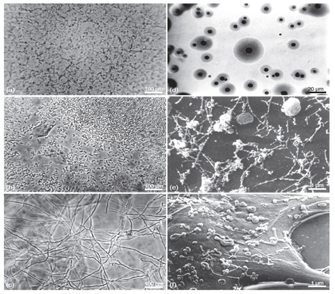
Figure-1: Types of Contamination. Examples of microorganisms found to contaminate cell cultures. (a) Bacteria. (b) Yeast. (c) Mold. (d) Mycoplasma colonies growing on special nutrient agar (e, f). Scanning electron micrograph of mycoplasma growing on the surface of cultured cells.