Michelson interferograms provide direct visual evidence of the mechanism of growth, e.g. spiral growth, two-dimensional nucleation, or birth-and-spread growth. Initially the interferometer is aligned with wedge fringes, which are then separated to yield the infinite-fringe interferogram. Under this setting the minute micro-morphological details become visible. They indicate the growth mechanism by which a particular face grows with time. These features are observed in-situ and in real time.
Sample images recorded in a crystal growth experiment by the Michelson interferometer are shown in Figure 4.69.
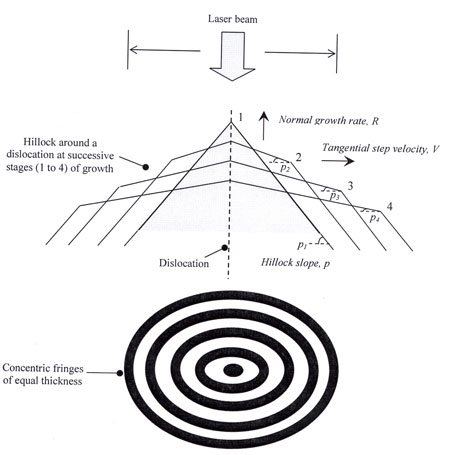
Figure 4.69: Schematic drawing of the Michelson Fringe formation from a crystal face having a hillock generated from a dislocation
|