Visualization of Band after Electrophoresis:
Most of the proteins are not colored. Thus, gel after electrophoresis has to be stained using protein specific dye. Coomassie dye (detection limit 200ng) is commonly used in laboratories for the purpose. The Coomassie dyes bind to proteins through ionic interactions between dye sulfonic acid groups and amine groups of basic amino acids as well as through Van der Waals interactions. As number of basic amino acid in a protein varies, this most commonly used dye don't stain all protein with equal affinity. Less
sensitive
dye include ponceau red (detection limit 250 ng) and amido black (detection limit 400 ng). The most sensitive staining is silver staining (detection limit 1 ng). This involves soaking the gel in AgNO3 which results in precipation of metallic silver (Ag0) at the location of protein forming a colored deposit (Fig. 3).
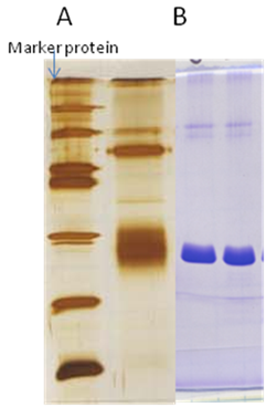
Figure 3: (A) gel stained with silver staining (B) gel stained with coomassie dye
|